



What are the Symptoms, Subtypes and Differential Diagnosis of Rosacea?
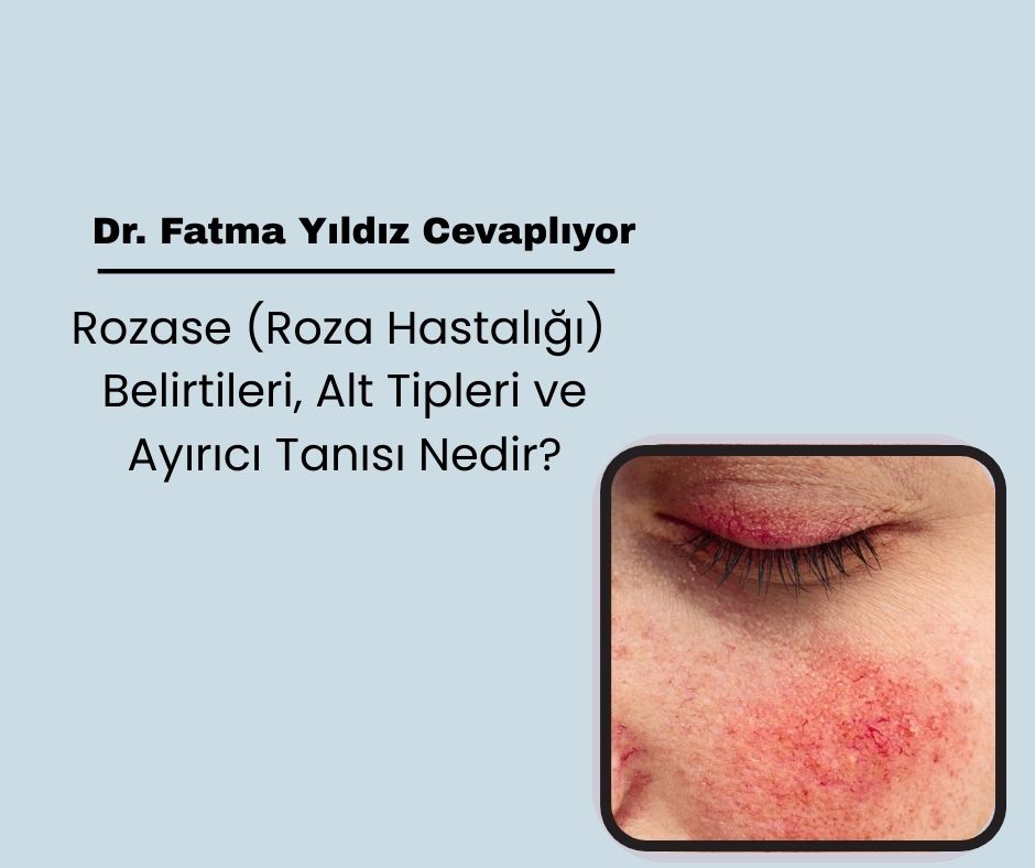
Rosacea is a chronic skin disease that is common in the adult age group. It manifests itself with inflammatory lesions (papules and pustules) accompanied by a burning and stinging sensation, especially in the middle part of the face, permanent redness (erythema), capillary dilatations (telangiectasia) and skin thickening (phymatous changes).
Four main subtypes of rosacea have been defined:
Erythematotelangiectatic rosacea
Papulopustular rosacea
Phymatous rosacea
Ocular rosacea
In addition to these, there are two rare variants:
Fulminant rosacea
Granulomatous rosacea
The same patient may show the findings of more than one subtype at the same time.
Clinical Findings in Rosacea
There are some factors that trigger redness attacks in rosacea patients. These include:
Emotional stress
Hot drinks
Spicy foods
Alcohol consumption
Exercise
Hot baths
These attacks are usually accompanied by a burning and stinging sensation. Patients' skin becomes more sensitive to cosmetic products. Sunscreens and some skin care products can cause discomfort such as itching and stinging.
Inflammatory papules and pustules are the distinctive symptoms of rosacea. However, unlike acne vulgaris, rosacea lesions do not contain comedones (blackheads). This helps distinguish rosacea from acne.
Over time, patients may develop yellow-orange plaques on the nose due to the enlargement of the sebaceous glands (rhinophyma).
Rosacea Appearance According to Race
In European and North American individuals, rosacea presents a more distinct picture in the form of red papules, pustules and telangiectasias. In contrast, redness is less noticeable in Asian, Latino and African individuals; Granulomatous type findings are more prominent. In African American patients, redness is less pronounced due to pigmentation, and hard, raised lesions are more common.
Classifications Used in Rosacea Diagnosis
A classification was developed by the National Rosacea Society (NRS) in 2002 to standardize the diagnosis of rosacea.
According to this classification, at least one of the primary findings must be present for diagnosis. Secondary findings support the diagnosis but are not mandatory.
Primary findings:
Redness attacks (flushing)
Persistent erythema
Papules and pustules
Telangiectasias
Secondary findings:
Burning, stinging sensation
Skin plaque formation
Dryness
Edema
Eye findings (ocular findings)
Spread to the face
Phymatous changes
Plewig-Kligman staging also defines the development of rosacea in stages:
Pre-rosacea: Frequent erythema attacks, increased sensitivity of the skin
Stage 1: Persistent redness and mild vascular dilatations
Stage 2: Addition of papules and pustules to persistent erythema
Stage 3: Large inflammatory nodules and skin thickening
Rosacea Subtypes
Erythematotelangiectatic Rosacea (Type 1)
In this subtype, long-lasting redness attacks and erythema that becomes permanent over time are seen in the central part of the face. Telangiectasias are a common finding. Burning and stinging sensations are prominent. Redness can sometimes spread to the outer areas of the face, ears and neck. There are mild, moderate and severe forms depending on the severity.
Papulopustular Rosacea (Type 2)
Small red blisters (papules) and pus-filled blisters (pustules) predominate on the central face. Burning and stinging sensations are less severe in this subtype. Chronic edema (swelling) may develop on the face in severe cases.
Phymatous Rosacea (Type 3)
In this form, thickening and irregular nodules are seen on the skin, especially around the nose (rhinophyma). Changes occur in the facial features due to the enlargement of the sebaceous glands. It is more common in men than in women. Phymatous changes can also occur on the forehead, chin, ears and eyelids.
Ocular Rosacea (Type 4)
Symptoms such as watering, stinging, burning, foreign body sensation, itching, light sensitivity and blurred vision are present in ocular rosacea. Redness (blepharitis) and conjunctivitis may also develop at the edges of the eyelids. In severe cases, there is a risk of corneal ulcers and vision loss.
Eye involvement develops in 50-60% of rosacea patients at some stage of the disease. There are also cases that start with eye complaints without skin symptoms.
Variants of Rosacea
Fulminant Rosacea (Pyoderma Fasciale)
A rapid onset and severe form, mostly seen in young women. Pustules, nodules and inflamed cysts that merge on the chin, cheeks and forehead occur. Comedones are usually not present. After the inflammatory period of the disease has passed, the classic rosacea picture may develop.
Granulomatous Rosacea
It presents with yellowish or brownish hard swellings on the thickened skin on the cheeks and around the mouth. Granulomas form in the subcutaneous tissue. Histological examination reveals structures that resemble sarcoidosis or lupus vulgaris. Granulomatous rosacea is more common in dark-skinned individuals.
Differential Diagnosis of Rosacea
Differential diagnosis is important because rosacea can be confused with some other diseases. These diseases include:
Acne vulgaris: Papules and pustules are present, but telangiectasia and flushing (facial redness) are absent as in rosacea. Comedones are also typical for acne.
Seborrheic dermatitis: There is scaling and eczematous changes on the face. The localization is usually the paranasal regions and behind the ears.
Perioral dermatitis: Smaller papules are limited to the area around the mouth, telangiectasia is absent.
Steroid rosacea: Develops after corticosteroid use and telangiectasia is prominent.
Contact dermatitis: Occurs as a result of contact with an irritant substance, usually with distinct borders.
Photodermatitis: Sun-related skin lesions are seen, usually affecting the areas of the body exposed to sunlight.
Lupus erythematosus: Butterfly-shaped redness is seen especially on the nose and cheeks, photosensitivity is high.
In addition, systemic diseases (such as polycythemia vera, mastocytosis, carcinoid syndrome, migraine) and some medications should be considered in the differential diagnosis as they can cause facial redness.
CONCLUSION
Rosa disease can present itself in many different clinical types and severities.
Correct recognition of the disease and differentiation from similar diseases are critical for appropriate treatment and management.
 Call
Call
 Whatsapp