


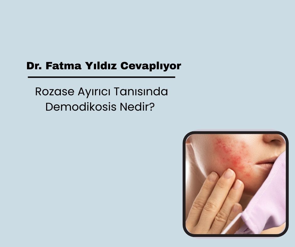

What is Demodicosis in the Differential Diagnosis of Rosacea?
Rosacea is a chronic facial dermatosis that is usually seen in middle-aged and older women and progresses with inflammatory attacks. It most commonly affects facial areas such as the forehead, nose, cheeks and chin. The disease initially begins with temporary erythema attacks (flushing) that develop with a feeling of warmth on the face — sometimes in the neck and chest area. Permanent erythema and telangiectasias develop over time. While the erythematous-telangiectatic type continues in this way, erythematous papules, pustules and rarely nodules can be added to the papulopustular type.
The pathogenesis of rosacea has not yet been fully elucidated. Various factors such as genetic predisposition, endocrine factors, vasomotor imbalance and Demodex spp. found in hair follicles are thought to be effective.
Demodicosis
Demodicosis is a chronic parasitic skin disease caused by Demodex mites of the Demodicidae family, usually affecting the face. It was first identified by Henle in 1841, and was shown to settle in pilosebaceous follicles by Simon in the same year. In 1934, Stokes and Beerman suggested that there may be a relationship between Demodex folliculorum and rosacea.
Demodicosis is the name given to dermatoses accompanied not only by an increase in Demodex mites but also by clinical findings related to this increase. Its incidence increases with age; it is rare in children, and this is attributed to low sebum production. The adult form of the mites is spindle-shaped, 250–400 µm long. Their bodies include a cephalothorax consisting of the head and thorax, an abdomen and 4 pairs of legs. Their life cycle is approximately 15 days and passes through hair follicles and sebaceous glands.
There are two types that are pathogenic in humans:
D. folliculorum: It is usually found in groups in the pilosebaceous ducts.
D. brevis: It lives alone in the sebaceous glands and Meibomian glands.
Pathogenesis
D. folliculorum mostly remains saprophytic and only causes immunological or allergic reactions in some individuals. The number of mites increases due to factors such as weakness in the immune system or increased sebum production. This situation may cause a non-inflammatory “pityriasis folliculorum” picture. Hyperkeratosis, which develops due to the increased number of mites, causes the follicle and sebaceous duct to become blocked and widened. With the rupture of the duct, the parasite spreads to the surrounding tissues and an inflammatory response begins. At this stage, erythematous papules, pustules and sometimes nodules may form.
Classification
Forton et al. (2014) suggested that “rosacea-like demodicosis” is the same disease as papulopustular rosacea and suggested subclassifications as “inflammatory/non-inflammatory” and “isolated/associated with other dermatoses”.
Chen et al. divided demodicosis into two main groups:
Primary Demodicosis: It is defined by the detection of mite increase in active lesions without another inflammatory dermatosis such as acne, rosacea or perioral dermatitis and only response to antiparasitic treatment. It is usually middle-aged and older. Asymmetric, irregularly shaped group lesions and satellite lesions are seen on the face.
Secondary Demodicosis: It develops in the background of another skin or systemic disease. It is especially seen in patients with immune deficiency (e.g. leukemia, HIV) and those using immunosuppressive drugs. It may be associated with dermatoses such as perioral dermatitis, seborrheic dermatitis, steroid rosacea. It may start at an earlier age than the primary type and is more commonly affected.
Clinical
The disease can be seen primarily on the face, scalp and anterior chest; rarely on the ears, back, breasts and genital areas. Symptoms include itching, burning, tenderness, dryness and coarsening of the skin.
Clinical findings:
Erythematous plaques, papules and pustules
Rarely nodulocystic lesions
Pityriasis folliculorum: Squamous lesions that feel like sandpaper
Demodex blepharitis: Dandruff at the eyelash roots
Demodex capitis: In the parieto-temporal alopecia area
Auricular demodicosis: Associated with chronic ear itching and external otitis
Differential Diagnosis
Rosacea
Demodicosis can mimic papulopustular rosacea in particular. Forton et al. showed that 74% of patients with papulopustular rosacea were positive for demodicosis in standard superficial skin biopsy. In addition, Demodex infestation causing granulomatous reaction can be confused with granulomatous rosacea. Demodex blepharitis can mimic ocular rosacea.
Distinguishing features:
Unlike rosacea, it usually occurs on one side of the face or asymmetrically.
Lesions are irregularly shaped, satellite lesions can be seen.
Inflammatory (papule/pustule) and non-inflammatory (pityriasis folliculorum) lesions can occur simultaneously.
Other Facial Dermatoses
Demodicosis can be seen together with seborrheic dermatitis, perioral dermatitis, contact dermatitis and atopic dermatitis. Especially a dominant picture such as seborrheic dermatitis can make the diagnosis of demodicosis difficult and may be a cause of resistance to treatment.
 Call
Call
 Whatsapp